Osteoporosis
You need clear, reliable information about osteoporosis, a condition where bones become weak and prone to fractures; this post explains what it is, its causes, risk factors, signs, differences from osteopenia and osteomalacia, diagnosis, diet and foods to avoid, prevention strategies, medical and homeopathic treatments, and natural remedies to support bone strength so you can make informed decisions about managing your bone health.
Key Takeaways:
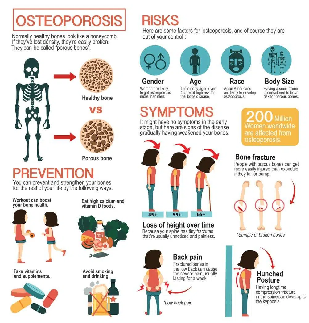
- Osteoporosis is a systemic skeletal disease characterized by low bone mass and microarchitectural deterioration, increasing fracture risk even with low trauma.
- Main causes and risk factors include aging, estrogen deficiency (postmenopause), genetic predisposition, low calcium/vitamin D, a sedentary lifestyle, smoking, excessive alcohol, low body weight, long-term glucocorticoid use, and certain chronic diseases.
- Osteoporosis often remains silent until it fractures; potential symptoms include back pain, height loss, stooped posture, and fractures from minor trauma.
- Diagnosis is determined using dual-energy X-ray absorptiometry (DXA) T-score, where osteopenia is defined as a score between −1.0 and −2.5, and osteoporosis is defined as a score of ≤ −2.5. Additionally, laboratory tests and imaging are conducted to identify any secondary causes; in contrast, osteomalacia is characterized by defective bone mineralization due to low vitamin D levels and elevated alkaline phosphatase.
- Diet and lifestyle: emphasize adequate calcium and vitamin D, regular weight-bearing and resistance exercise, sufficient protein, and fruits/vegetables; avoid high-salt diets, excessive caffeine, soda/phosphoric acid beverages, heavy alcohol use, and smoking.
- Treatment options include antiresorptives (bisphosphonates, denosumab), anabolic agents (teriparatide, romosozumab), SERMs and others, as indicated, plus calcium/vitamin D supplementation and fall-prevention measures.
- Natural approaches (exercise, nutrition, and fall prevention) support bone health; homeopathic remedies lack reliable evidence as effective treatments and should not replace proven medical therapy—discuss any complementary use with a healthcare provider.
The Anatomy of Osteoporosis: What You Need to Know
Defining Osteoporosis: The Silent Crippler
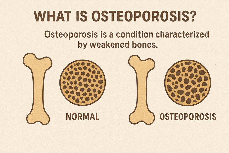
You accumulate most of your peak bone mass by your late 20s—genetics accounts for roughly 60–80% of that peak—so what happens after that largely determines whether you’ll develop osteoporosis. The condition represents a progressive loss of bone mass and deterioration of bone microarchitecture: trabeculae thin and disconnect, cortical bone becomes porous, and overall strength drops even if you don’t notice symptoms. Clinical definitions rely on bone mineral density (BMD) measurements: a T‑score between -1.0 and -2.5 is labeled osteopenia, and a T‑score ≤ -2.5 defines osteoporosis; however, those numbers are only part of the story because structural integrity and fall risk also shape your fracture risk.
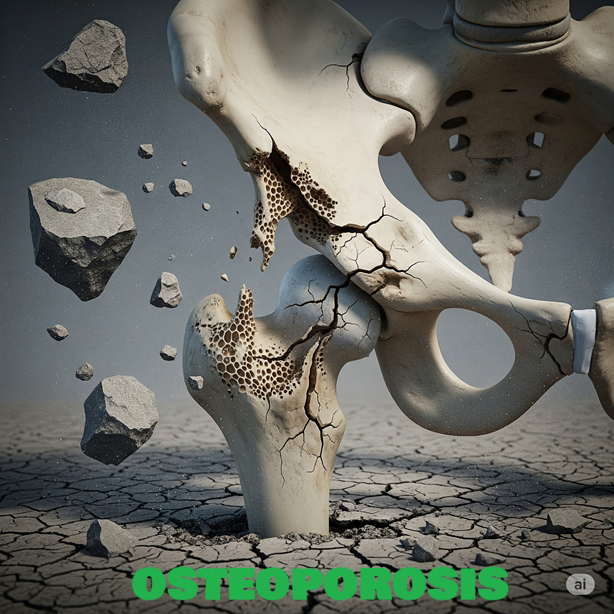
You often won’t feel any change until a fracture occurs, which is why osteoporosis earns the label “silent” in practice. Vertebral compression fractures can happen with minimal trauma and may present only as gradual height loss (sometimes 2–6 cm over years) or a stooped posture; hip and distal radius fractures tend to be the first events that bring people into acute care. Epidemiologic data underline the stakes: about 1 in 3 women and 1 in 5 men over 50 will sustain an osteoporosis-related fracture in their remaining lifetime, and hip fractures carry a 20–30% increased mortality in the year following injury. Given that you might show no pain until structural failure occurs, screening and awareness of risk factors—postmenopausal estrogen decline, long-term glucocorticoid use, low BMI, smoking, heavy alcohol intake, and malabsorption syndromes—are relevant for early detection.
You can trace many cases back to two interacting processes: insufficient peak bone mass and accelerated bone loss. Peak bone mass is largely set by age 30; thereafter, low-level bone remodeling balances formation and resorption until factors like menopause tilt the balance toward resorption. Postmenopausal women typically experience 1–2% annual bone loss, with more rapid losses (up to 3–5% per year) in the early postmenopausal years for some; men lose bone more slowly but increasingly after age 70. Clinical vignettes illustrate this pattern: a 68‑year‑old woman with a femoral neck T‑score of -2.8 may present after a low‑impact fall with a hip fracture, whereas a 60‑year‑old man with the same T‑score might remain fracture-free for years—differences that reflect both time course and additional risk modifiers like balance, vision, and medication use that you must consider alongside BMD.
Bone Density and Structural Integrity: Why It Matters
You measure bone density with dual-energy X‑ray absorptiometry (DEXA), which gives the T‑score used for diagnosis, but density alone underestimates the complexity of fracture risk. Microarchitectural features—trabecular thickness and number, connectivity, cortical thickness and porosity—determine how your bone distributes loads and resists crack propagation. Advanced imaging techniques like high-resolution peripheral quantitative CT (HR‑pQCT) and finite element analysis can show problems in trabecular connectivity or cortical porosity that DEXA misses, which is why two people with the same bone mineral density (BMD) can
You also need to factor in bone turnover dynamics: markers like serum C‑telopeptide (CTX) for resorption and P1NP for formation reflect the velocity of remodeling. High turnover states—induced by hyperthyroidism, prolonged glucocorticoid therapy, or inflammatory diseases—erode microarchitecture faster than BMD changes might suggest, leaving you at elevated fracture risk even with modest T‑score reductions. Doctors are now considering these markers and imaging more in their decisions: a patient taking long-term steroids with nearly normal BMD but very high CTX levels might receive more aggressive treatment than a patient with low turnover who has the same T-score
You can quantify the impact: each 1 standard deviation decrease in femoral neck BMD approximately doubles your risk of hip fracture, and the cumulative effect of microarchitectural loss plus lower BMD multiplies that risk further. Real-life examples highlight the difference—someone with a T-score of -2.0 and healthy trabecular structure might have a lower chance of breaking a bone in the next 10 years compared to someone with a T-score of -1.8 but serious trabecular damage and a history of falls—so evaluations that consider BMD, bone quality, fall history, and other health factors give the best understanding of your fracture
Triggers of Bone Loss: Unpacking the Causes
Hormonal Changes: Menopause and Beyond
Estrogen acts directly on osteoclasts and osteoblasts, and after menopause your estrogen levels typically fall sharply, producing bone loss rates that can reach about 2–3% per year in the first 5–7 years; cumulative loss in that window can approach 10% of total bone mass. Men experience more gradual hormonal decline—testosterone and estradiol reductions usually start later, and you generally lose bone at roughly 1% per year after age 60—yet hypogonadism at any age accelerates osteoporosis risk. If you are treated with endocrine therapies (for example, aromatase inhibitors for breast cancer) or if you have had surgical menopause, expect a faster trajectory of bone density decline and a higher fracture risk unless countermeasures are in place.
Corticosteroid-induced osteoporosis is one of the most predictable medication-related causes: taking prednisone at doses as low as 5 mg per day for three months or longer raises your fracture risk significantly, and bone loss can begin within weeks of starting therapy. Thyroid hormone excess from overtreatment or uncontrolled hyperthyroidism increases bone turnover and lowers BMD, particularly in cortical bone such as the hip; subclinical hyperthyroidism also carries measurable risk. Primary hyperparathyroidism and other endocrine disorders (Cushing’s syndrome, uncontrolled diabetes) alter calcium handling and bone remodeling, so you should screen for endocrine causes if bone loss appears rapid or out of proportion to age.
Genetic and developmental hormonal influences also matter: early menopause (before age 45), amenorrhea from athletic training or eating disorders, and childhood conditions that impair peak bone mass all set the stage for earlier osteoporosis. You can measure fracture risk more accurately by combining hormonal history with DEXA results and tools like FRAX; for example, a woman with menopause at 42 and a T-score of -2.0 will have a higher 10-year fracture probability than a peer who entered menopause at 52 with the same T-score. When you identify endocrine triggers, your management plan should include targeted hormonal evaluation and intervention—ranging from minimizing glucocorticoid exposure to considering bone-protective drugs in patients on aromatase inhibitors.
Nutritional Deficiencies: The Role of Calcium and Vitamin D
Calcium is the structural scaffold of bone, and if your dietary intake falls short over years, bone remodeling will draw on skeletal reserves. Most adults need roughly 1,000–1,200 mg of elemental calcium daily from food and supplements combined; studies show that populations consuming less than 700 mg/day have higher osteoporotic fracture rates. You absorb dietary calcium less efficiently with age and in the presence of low gastric acid or after bariatric surgery, and conditions like celiac disease or inflammatory bowel disease can reduce absorption dramatically, producing secondary bone loss.
Vitamin D governs intestinal calcium absorption and has direct effects on muscle function and fall risk; deficiency is linked to lower BMD and higher fracture incidence. For older adults, many guidelines support a target intake of about 800–1,000 IU/day to maintain 25(OH)D levels above 20–30 ng/mL, though individuals with obesity, malabsorption, or darker skin pigmentation often require higher supplementation to reach target levels. Seasonal studies demonstrate that populations in higher latitudes commonly show winter nadirs in 25(OH)D and corresponding small but measurable falls in calcium absorption and bone turnover markers.
Not getting enough protein can hurt the formation of bone structure, and lacking magnesium, vitamin K, and trace elements like zinc and copper has been linked to weaker bones in studies. Diets that are heavily skewed toward processed foods, have sodium levels over 2,300 mg/day, and follow high-phytate patterns without adequate calcium can reduce net calcium balance. If you don’t get enough nutrients from your diet or if your body has trouble absorbing them, you should consider taking supplements and changing your diet, based on tests for vitamin D, calcium, and bone health markers, to give your
More granularly, timing and form of supplementation matter: calcium carbonate requires an acidic stomach and is best taken with meals, whereas calcium citrate can be better absorbed in achlorhydric states; dividing doses above 500 mg optimizes absorption. Measuring serum 25(OH)D rather than relying on sun exposure estimates lets you titrate vitamin D to a therapeutic range; in practice, loading doses (for example, 50,000 IU weekly for 6–8 weeks) followed by maintenance have been used to correct deficiency under clinical supervision. Combining adequate calcium and vitamin D with weight-bearing exercise produces synergistic benefits, and you should avoid excess calcium supplementation without monitoring because of potential cardiovascular and renal stone risks in susceptible individuals.
Lifestyle Factors: Smoking, Alcohol, and Physical Activity
Tobacco use speeds up bone loss by harming bone-building cells, changing how sex hormones work, and lowering calcium absorption in the gut; many studies show that current smokers are 30–40% more likely to have a hip fracture than those who have never smoked.
Nicotine and other components impair fracture healing as well, so if you smoke and sustain a low-trauma fracture, expect slower recovery and higher complication rates. Quitting reduces risk over time, but the bone density advantages accumulate gradually, so cessation is a long-term investment in your skeletal health.
Alcohol has a dose-dependent relationship with bone: light-to-moderate intake may have minimal effects, but habitual consumption above two standard drinks per day increases fracture risk and interferes with osteoblast function and calcium balance. Epidemiological data link heavy drinking (for example, more than three drinks daily) to both falls—through impaired coordination—and poorer BMD. Your pattern of consumption matters; binge drinking events and chronic excess carry different but additive risks for bone strength and fall likelihood.
Being active is one of the most important things you can change to help your bones throughout your life: activities that put weight on your bones, like jogging and jumping, along with strength training, can help build new bone and increase bone mineral density (BMD) by 1–3% in 6–12 months in Conversely, prolonged immobility produces rapid losses—for instance, astronauts in microgravity can lose ~1–2% of bone mass per month, and patients on long bedrest show accelerated cortical thinning within weeks. Balance and functional training reduce fall risk independently of BMD, so your exercise program should combine resistance, impact-loading (as tolerated), and neuromuscular work to maximize fracture prevention.
- Smoking increases fracture risk and slows healing.
- Excess alcohol (>2 drinks/day) impairs bone formation and increases falls.
- Recognizing that a tailored exercise regimen (progressive resistance plus balance) yields both BMD gains and fewer falls helps you prioritize interventions.
Other lifestyle contributors—poor sleep, chronic stress with elevated cortisol, and certain dietary patterns high in sodium and low in protein—compound these risks by altering bone turnover and fall propensity; for example, high dietary sodium (more than 3,000 mg/day) increases urinary calcium losses and can worsen negative calcium balance. Combining smoking cessation, limiting alcohol, and a structured exercise plan produces multiplicative benefits: clinical programs that add resistance training to standard care report fewer incident fractures over time than lifestyle counseling alone. Understanding that combining multiple lifestyle changes usually provides a greater reduction in fracture risk than relying on individual measures will help inform your practical therapy choices.
- Address sleep, stress, and high-sodium diets to limit urinary calcium loss.
- Include resistance and balance training in rehabilitation and prevention plans.
- Recognizing the cumulative effect of multiple small lifestyle changes on long-term bone health is important for sustainable risk reduction.
Identifying the Risk: Are You at an Elevated Risk for Osteoporosis?
Genetic Factors: Family History and Ethnicity
Family history strongly influences your peak bone mass and fracture susceptibility; twin and family studies estimate heritability of bone mineral density between roughly 50% and 85%, so if your mother or father sustained a hip fracture, you carry a substantially higher baseline risk. Specific genetic variants—for example, polymorphisms in the COL1A1 gene (type I collagen), LRP5 (Wnt signaling), the vitamin D receptor, and estrogen receptor loci—have been associated with lower bone density and higher fracture rates in population studies and should be considered when you build your personal risk profile. Clinical examples show that two sisters with similar lifestyles can have markedly different DXA results because of inherited differences in bone geometry and microarchitecture, not just density.
- First‑degree relative with hip or vertebral fracture
- Known familial bone disorders (osteogenesis imperfecta, juvenile osteoporosis)
- Ethnic background: higher baseline risk among white (Northern European) and East Asian populations
Genetic testing for common polymorphisms is not routine in most osteoporosis clinics, but a detailed three‑generation family history will guide earlier screening and preventive steps; you should inform your clinician if multiple relatives had low‑energy fractures or early osteoporosis diagnoses. Lifestyle and medical care can greatly reduce the risk passed down in families—getting enough calcium (1,000–1,200 mg per day based on age), ensuring good vitamin D levels (aim for 25‑OH vitamin D of at least 30 ng/mL for many high The more affected first‑degree relatives you have with osteoporotic fractures, the higher your lifetime fracture probability and the earlier you should seek bone density testing.
Age and Gender: Who Is Most Vulnerable?
Women represent the majority of osteoporosis cases due to the significant decline in estrogen levels after menopause, which serves as a critical risk period. Estrogen withdrawal accelerates bone remodeling imbalance—resorption outpaces formation—and you can lose as much as 20% of trabecular bone mass in the first 5–7 years after menopause in some women, which explains the sharp rise in vertebral and wrist fractures in the sixth decade. If you experienced surgical menopause (bilateral oophorectomy) or primary ovarian insufficiency before age 45, your fracture risk mirrors that of older postmenopausal women unless you receive appropriate hormonal or bone‑protective interventions.
Men lose bone more gradually but are often diagnosed later because screening thresholds are higher and fractures occur at older ages; hypogonadism, long-term corticosteroid use, alcohol abuse, and smoking accelerate male bone loss. You should ask for a DXA scan if you have these concurrent r70, your absolute fracture risk rises for both sexes because of cumulative bone loss, reduced neuromuscular function, and areduced neuromuscular function, and higher fall frequency—hip fracture rates climb steeply in the eighth decade and beyond, with associated morbidity and mortality. Clinical practice patterns recommend earlier assessment when you have multiple risk factors: for example, a 58‑year‑old woman with early menopause and chronic steroid therapy warrants the same attention as an older patient without those issues.
Screening guidelines usually suggest getting a DXA scan for women 65 and older and men 70 and older, but if you have extra risk factors like a parent who had a hip fracture, a previous low-impact fracture, or long-term use of glucocorticoids, you should think about getting evaluated sooner; use the FRAX tool to combine factors like age, sex, weight, smoking, and family history of fractures to estimate your 10-year risk, which can A T‑score ≤ −2.5 defines osteoporosis, while −1.0 to −2.5 indicates low bone mass (osteopenia) and a different management threshold; if you fall into osteopenia but have high FRAX probability or prior fragility fracture, Additionally, if you are a man with hypogonadism, a breast cancer survivor taking aromatase inhibitors, or a woman who experienced prolonged amenorrhea in her younger years, your age-related baseline for bone health screening shifts earlier, and you should take proactive measures to maintain bone health.
Medical Conditions and Medications: The Hidden Dangers
Many health issues can lead to secondary osteoporosis by changing how bones are built, how nutrients are absorbed, or hormone levels; for example, hyperthyroidism speeds up bone loss and increases the chance of fractures, primary hyperparathyroidism causes loss of cortical bone, and long-term inflammatory diseases like rheumatoid arthritis or inflammatory bowel disease boost bone breakdown through pathways. Malabsorptive conditions like celiac disease and chronic pancreatitis reduce calcium and vitamin D uptake, while chronic kidney disease disrupts mineral homeostasis and active vitamin D synthesis, all of which lower your bone strength. Hormonal diseases—like untreated type 1 diabetes, low testosterone in men, and long periods without menstruation in women—make the risk even higher, so it’s important to check your medical history and do tests (like TSH, PTH, and 25‑OH vitamin) if you present with low bone density.
Medications often play a significant role in bone loss that can be changed: systemic glucocorticoids are the most common cause of drug-related bone loss, with effects that depend on the dose and how long you take them—taking doses similar to prednisone of 5 mg/day or more for three months or longer increases. Other medications that can increase the risk of fractures include aromatase inhibitors for hormone-sensitive breast cancer, androgen-deprivation therapy for prostate cancer, certain anticonvulsants like phenytoin and carbamazepine that affect vitamin D, selective serotonin reuptake inhibitors, and long-term use of proton pump inhibitors. If you take any of these drugs, discuss alternatives, bone‑protective co‑therapies, or surveillance plans with your prescriber so you don’t discover loss only after a fracture.
You can lower the risk from medications by regularly reviewing your prescriptions and keeping an eye on your health: get a baseline DXA scan before starting long-term glucocorticoid or aromatase inhibitor therapy, have follow-up scans based on your risk level, and check for other Practical steps include lowering the amount and time you take steroids, making sure you get enough calcium and vitamin D, and starting antiresorptive therapy (bisphosphonates or denosumab) for patients on long-term high-dose glucocorticoids or with existing osteoporosis; working together with your primary care doctor, rheumatologist or oncologist, and a
If you have a long-term bone disease, keep track of when you started taking high-risk medications and how long you’ve been on them, get a DXA scan and fall-risk assessment early, and talk about preventive medications—teriparatide or romosozumab might be better for very high-risk cases or those caused by glucocorticoids, while bisphosphonates are usually the first choice for most others; your treatment
Nutritional Pitfalls: Foods to Avoid If You Have Osteoporosis
High-Sodium Choices: The Link to Bone Loss
High-sodium processed foods force your kidneys to dump more calcium into the urine; clinical physiology shows that an extra 100 mmol of sodium (about 2,300 mg) corresponds roughly to an additional 1 mmol (≈40 mg) of urinary calcium loss. Packaged soups, canned vegetables, deli meats and many frozen dinners often contain 800–1,500 mg of sodium perving, so a single restaurant meal or convenience meal can push you well beyond your daily limits and measurably increase calcium excretion for the remainder of the day. Consistently elevated sodium intake accelerates negative calcium balance, and over months and years, that shift contributes to greater bone resorption and lower bone mineral density, particularly in postmenopausal women and older adults whose baseline calcium turnover is already higher.
When you compare dietary patterns, populations with lower average sodium intakes typically show slower rates of age-related bone loss; epidemiological data link high-salt diets to higher markers of bone turnover. Practical examples include swapping canned beans for rinsed canned or, better, cooked-from-dry legumes—rinsing can reduce sodium by up to 40%—or choosing low-sodium broths and seasoning with herbs and acids rather than salt. Grocery labels matter: products labeled “reduced sodium” still may contain 300–600 mg per serving, so keeping your daily target closer to 1,500 mg, or at least under 2,300 mg, will give you a tangible reduction in calcium wasting and lessen long-term bone loss risk.
Cooking strategy influences sodium exposure and bone outcomes: preparing meals from scratch, freezing single portions of low-sodium stews, and using citrus, garlic, and smoked paprika for flavor reduces reliance on salt without sacrificing palatability. Restaurant choices can be guarded by asking for sauces and dressings on the side—many entrées add 500–1,000 mg in a single portion of sauce—and choosing grilled options over heavily brined or cured proteins. Tracking sodium intake for a week using a food app will quickly show you the top culprits in your diet so you can target swaps that protect calcium balance and reduce the biochemical drivers of bone loss.
Caffeine and Alcohol: Moderation Is Key
Daily caffeine consumption above roughly 300 mg—about three 8-ounce cups of brewed coffee—has been associated in several studies with increased urinary calcium loss and a modestly higher risk of fractures in people who do not meet their calcium needs; if your calcium intake is below 800 mg/day, the effect is more pronounced. Coffee, certain teas, chocolate, and different energy drinks all deliver variable doses of caffeine, and the timing matters: consuming large amounts of caffeine around the time you take calcium-rich meals or supplements can reduce net calcium retention. You can still enjoy coffee, but spacing it at least one hour away from calcium supplements and aiming for no more than two to three standard cups a day will blunt most of the negative effects on calcium balance.
Alcohol interacts with bone through several mechanisms: heavy, chronic drinking (commonly defined as more than two standard drinks per day for men and more than one for women) impairs osteoblast function, disrupts vitamin D metabolism, and increases fall risk through sedation and impaired coordination—each pathway raises fracture risk. Binge drinking episodes that occur in a short time are particularly harmful because they combine metabolic effects with increased likelihood of falls; even moderate habitual drinking can reduce bone formation markers if combined with poor nutrition. For bone health, prioritize limiting alcohol to occasional, low-volume consumption and avoid using alcohol as a stress or sleep aid, since that pattern compounds risks to bone remodeling and muscular stability.
Standard drink definitions help you control intake: one standard drink equals 14 g of pure alcohol (12 oz beer at ~5%, 5 oz wine at ~12%, or 1.5 oz distilled spirits at ~40%), so tracking these units keeps you aware of cumulative exposure. If you take bisphosphonates or other osteoporosis medications, coordinate timing and avoid alcohol immediately around dosing and when you know booze will impair your balance; discuss specific interactions with your clinician if you consume it regularly. Swapping a late-night cocktail for herbal tea or a decaffeinated beverage reduces both fall risk and the metabolic burden on bone while preserving social habits.
Certain Foods That Can Inhibit Calcium Absorption
High-oxalate vegetables such as spinach, beet greens and rhubarb contain oxalic acid that binds calcium into insoluble complexes, making the calcium in those foods poorly bioavailable despite high total calcium content on paper. For example, a cup of raw spinach contains around 30–50 mg of calcium, but much of that calcium is locked up by oxalate, so absorption is far lower than from low-oxalate greens like kale or bok choy; choosing those lower-oxalate alternatives gives you more absorbable calcium per serving. Eating spinach in large quantities as your primary calcium source can therefore give you a false sense of security—if you rely on plant sources, prioritize low-oxalate choices and diversify with fortified foods or dairy if you tolerate it.
Phytic acids in whole grains, seeds, and legumes can chelate calcium and other minerals, reducing absorption when meals are heavily grain- or legume-based and unprocessed. Traditional food preparation methods—soaking, fermenting, sprouting and sourdough fermentation—reduce phytate levels substantially; for instance, sourdough bread and sprouted grain products have significantly higher mineral bioavailability than standard whole wheat bread. You can keep beans and lentils in your rotation by soaking and rinsing them before cooking or you can choose canned varieties that you can rinse, and include vitamin C-rich foods in the same meal to offset phytate effects and boost mineral uptake.
Fat malabsorption syndromes and very high-fiber diets can also make it harder for your body to absorb calcium: when unabsorbed fatty acids attach to calcium and form insoluble soaps, less calcium is available for your body to use, which can happen in conditions like untreated celiac disease or Tannins in strong black tea and some herbal blends reduce mineral uptake when consumed in large amounts with meals; for example, sipping multiple cups of strong tea with a high-calcium meal will blunt absorption compared with drinking water. Pair calcium-rich foods with vitamin D sources and acidic foods like citrus to enhance uptake, and space high-tannin beverages away from meals if you rely on dietary calcium.
Choose calcium sources strategically: if you want plant-based options with good bioavailability, go for kale, bok choy, broccoli and fortified plant milks; one cup of cooked kale can supply roughly 100–180 mg of well-absorbed calcium depending on preparation. When you include grains and legumes, apply soaking, sprouting or fermentation to lower phytate content, and combine those dishes with vitamin C–rich sides (peppers, citrus, kiwi) to improve absorption. If you take calcium supplements, avoid taking them at the same time as high-oxalate meals or iron supplements—spacing doses by one to two hours maximizes net absorption and minimizes competitive inhibition.
Nourishing Your Bones: A Dietary Blueprint for Osteoporosis
Foods to Embrace: Calcium and Protein Powerhouses
Aim for dietary calcium sources that give you both high bioavailability and predictable portion values: a standard 8‑oz (240‑ml) cup of cow’s milk supplies roughly 300 mg of calcium, the same ballpark as 1 cup of fortified plant milk or ¾ cup of low‑fat yogurt. Tofu prepared with calcium sulfate can contribute 200–350 mg per half‑cup depending on the product, and a 3‑oz (85‑g) serving of canned salmon with bones typically supplies around 150–200 mg. Dark leafy greens such as cooked kale and bok choy offer absorbable calcium (about 90–100 mg per cooked cup), whereas spinach and Swiss chard contain similar total calcium but are high in oxalates that sharply reduce absorption—so you should not rely on those as primary calcium sources.
Prioritize protein at each meal because amino acids support bone remodeling and preserve muscle. Older adults should aim for roughly 1.0–1.2 g of protein per kg of body weight each day; for example, a person weighing 70 kg would target 70–84 g of protein daily. Practical sources include lean poultry, fish, eggs, dairy, legumes, and soy; a single 3‑oz (85‑g) portion of cooked salmon or chicken is typically 20–25 g of protein, a cup of cooked lentils provides ~18 g, and a cup of Greek yogurt gives 15–20 g. Distribute protein evenly—shooting for 20–30 g per meal—because trials and cohort studies show better preservation of hip and spine bone mineral density when protein intake is adequate and evenly spaced.
Pair calcium and protein intentionally in meals while keeping sodium and caffeine in check to limit urinary calcium losses; follow the general dietary sodium recommendation of under 2,300 mg/day to reduce calcium excretion. Practical meal patterns include a breakfast of fortified oat milk with high‑protein cereal and a dollop of Greek yogurt (≈300 mg calcium + 20 g protein), a lunch of sautéed tofu and bok choy over brown rice (≈250 mg calcium + 20–25 g protein), and a dinner of canned salmon salad with mixed greens and a side of lentils (≈200 mg calcium + 25–30 g protein). You can use fortified foods to plug gaps—many plant milks and breakfast cereals contain ~300 mg calcium per serving—so tally what you eat across a day to reach the 1,000–1,200 mg total calcium target recommended for most adults (1,000 mg for men and women under 50; 1,200 mg for women over 50 and men over 70).
The Role of Supplements: When Dietary Adjustments Are Not Enough
If your dietary intake consistently falls well below targets—for example, you average under ~700 mg calcium per day—or if you have malabsorption (celiac disease, post‑gastric bypass), documented vitamin D deficiency, or strict avoidance of dairy, supplements become a practical tool. Choose a calcium formulation based on tolerance and acid status: calcium carbonate provides about 40% elemental calcium and is cost‑effective but requires food for optimal absorption; calcium citrate contains ~21% elemental calcium, is better absorbed with low stomach acid or proton‑pump inhibitor use, and can be taken with or without food. Split supplemental calcium into doses no greater than 500 mg elemental calcium at a time to maximize absorption, since absorption efficiency drops above that dose.
Target vitamin D to support intestinal calcium uptake and muscle function by checking a serum 25‑hydroxyvitamin D level before initiating high‑dose therapy; many clinicians aim for a 25(OH)D ≥30 ng/mL for bone health. Typical maintenance dosing ranges from 800 to 2,000 IU of cholecalciferol (D3) daily for older adults, with higher or loading regimens used under supervision for deficiency—retest after 8–12 weeks of supplementation. Be mindful of interactions and timing: Separate calcium supplements from oral bisphosphonates, levothyroxine, and certain fluoroquinolone or tetracycline antibiotics to prevent reduced drug absorption. Take bisphosphonates according to label instructions on an empty stomach, and delay taking calcium for at least 30–60 minutes or as recommended.
Consider additional micronutrients that influence bone metabolism where dietary intake or lab testing shows gaps: Magnesium (recommended amounts are about 310–420 mg per day based on your sex and age) helps activate vitamin D and control parathyroid hormone; also, vitamin K2 (MK-7), usually taken at 90–200 mcg per day in supplements, may help move calcium into bones instead of soft tissue, but be careful if you are on warfarin or other blood thinners. g/day in trials) combined with vitamin C have produced modest improvements in some bone markers and BMD in small studies, though evidence is not yet definitive. Coordinate supplementation with your clinician: measure baseline labs (25[OH]D, serum calcium, creatinine), set a target plan (for instance, add 600 mg elemental calcium and 1,000–2,000 IU vitamin D3 daily for someone with dietary calcium ~600 mg and low vitamin D), and recheck labs in 3 months to adjust dosing; monitor urinary calcium if you have a history of nephrolithiasis; and avoid exceeding combined dietary and supplemental calcium routinely above 1,500–2,000 mg/day without specialist oversight.
Recognizing the Signs: Symptoms that Signal Osteoporosis
Early Warning Signs: What to Watch For
You may first notice subtle back pain that seems to come from your mid- or lower thoracic spine rather than from muscle strain; studies show vertebral compression fractures can be painless or present as chronic, nagging mid‑back discomfort that worsens with activity and improves with rest. A common objective clue is loss of height—losing 2 cm (about 0.8 inch) or more over a year should prompt evaluation, since single or multiple wedge fractures of the vertebrae often produce measurable height loss. In clinical series, patients with unrecognized vertebral fractures frequently report progressive stooping or a change in posture that they attribute to “aging,” yet that kyphotic change often reflects underlying bone loss.
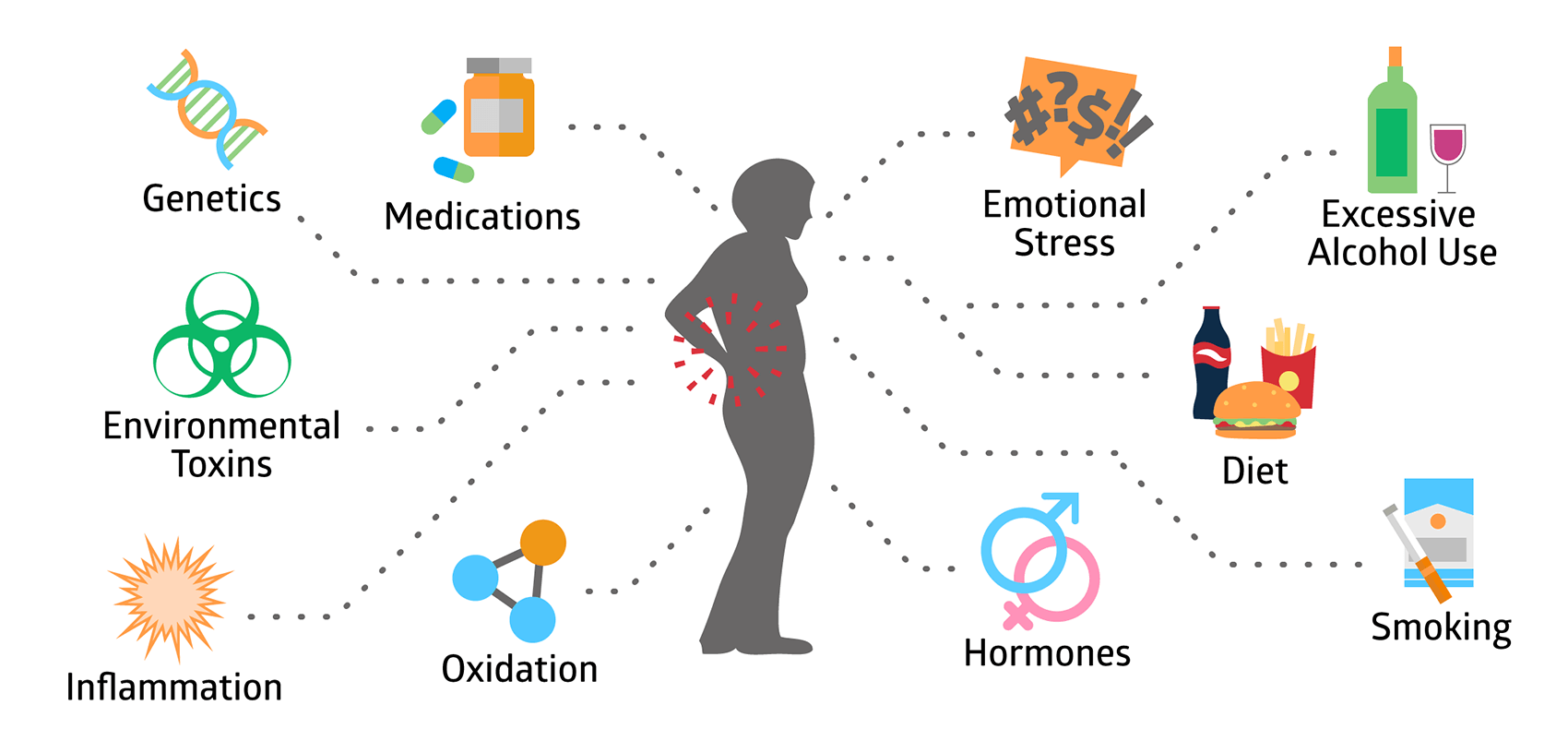
You might notice fractures from low-impact events, such as slipping and landing on a hip or breaking a wrist after a fall from standing height, may be noticeable because healthy bone typically withstands these forces. Bone mineral density (BMD) tests show that osteopenia (a T-score between −1.0 and −2.5) can turn into osteoporosis (a T-score of −2.5 or lower) over a few years, which means that having one low-impact fracture in your 50s or glucocorticoid use (>3 months), smoking, alcohol intake >3 units/day, early menopause, or a family history of hip fracture, a single fragility fracture should trigger prompt assessment and possible treatment.
You may experience recurrent minor fractures or persistent, poorly localized pain that does not respond to typical musculoskeletal therapies; case reports often describe patients treated for “mechanical back pain” for months before spinal radiographs reveal multiple compression fractures. Quantitative details help: a single vertebral compression can reduce vertebral height by 15–30%, and multiple compressed vertebrae compound functional decline and pain. If you notice multiple small fractures or increasing reliance on analgesics to function, that pattern typically signals that bone loss is advanced enough to require medical intervention rather than conservative therapy alone.
- New, unexplained back pain persisting >6 weeks with postural change
- Height loss ≥2 cm over 12 months or progressive stoop
- Low‑impact fractures (wrist, hip, vertebra) after a fall from standing
- Long‑term steroid therapy or other secondary osteoporosis risk factors
- Repeated fractures or persistent pain despite standard musculoskeletal treatment
Early Signs: Quick Reference
| Sign | Why it matters |
|---|---|
| New mid‑back pain | May indicate vertebral compression fracture reducing vertebral height and spinal stability |
| Height loss ≥2 cm | Objective measure suggesting one or more vertebral fractures or progressive bone loss |
| Low‑impact fracture | Signals diminished bone strength; often the first clinically evident event |
| Risk factor cluster (steroids, smoking, low BMI) | Accelerates bone loss and elevates short‑term fracture risk |
Advanced Symptoms: When to Seek Help
You will notice more pronounced functional impairment as osteoporosis progresses: inability to rise from a chair without using your arms, difficulty with activities that were previously effortless, and persistent pain that interferes with sleep and daily tasks. Hip fractures from a simple fall often lead to hospitalization and surgery; epidemiologic data show roughly one in five men and one in three women over 50 will sustain an osteoporotic fracture in their lifetime, and hip fractures in particular carry significant morbidity—many patients require long‑term care or lose independent living. If routine activities like climbing a single flight of stairs become painful or unstable, that level of decline often reflects advanced bone fragility or accumulated fractures.
Neurological symptoms can emerge when several vertebrae in the middle or lower back collapse; it can cause nerve problems, like pain, numbness, or weakness in the legs, which may mean that broken bone pieces are pressing on nerves. Imaging tests often show that untreated vertebral compression can harm lung function and cause ongoing pain; for instance, having multiple front wedge fractures can reduce chest space and make it hard to take deep breaths. Sudden, severe back pain along with new problems with bowel, bladder, or leg function needs to be checked right away because it could mean a serious spine injury or pressure on the spinal cord.
The risk of experiencing another fracture in the year following an initial vertebral fracture increases dramatically; some studies indicate a two- to five-fold rise in short-term fracture risk. Persistent structural deformity, chronic opioid‑dependent pain, and loss of height greater than 5 cm often indicate longstanding, untreated osteoporosis with cumulative skeletal compromise. You should act quickly if you experience recurrent fractures or escalating functional loss, as timely intervention can reduce further fracture risk and improve quality of life.
Advanced symptoms often signal a need Serious symptoms usually mean you need imaging tests (like spine X-rays or a DEXA scan) and help from different specialists; if a vertebra collapses and leads to a hunched back or nerve issues, you might need surgery along with
- Neurologic signs (numbness, weakness)
- Hip or pelvic fracture after a low‑energy fall
- Marked height loss >5 cm or progressive kyphosis
- Repeated fractures within 12–24 months
- Loss of independence in activities of daily living
Advanced Signs: Clinical Details
| Sign | Clinical implication |
|---|---|
| Hip fracture from standing fall | Often requires surgical fixation; high risk of postoperative morbidity and loss of independence |
| Multiple vertebral compressions | Leads to chronic pain, deformity, reduced pulmonary capacity, and increased future fracture risk |
| Neurologic deficits | May indicate neural compression from bone fragments—urgent imaging and referral necessary |
| Rapid fracture recurrence | Suggests very high short‑term fracture risk and a need for immediate antiresorptive or anabolic therapy |
Differentiating Bone Conditions: Osteopenia vs. Osteoporosis vs. Osteomalacia
Key Differences: Understanding the Distinctions
Osteopenia and osteoporosis are defined by bone mineral density (BMD) measured on DXA: osteopenia corresponds to a T-score between -1.0 and -2.5, while osteoporosis is a T-score of -2.5 or lower. You should know that each standard deviation drop in BMD roughly doubles your fracture risk, which helps explain why a T-score crossing the -2.5 threshold changes management from preventive measures to active pharmacologic therapy. Epidemiologically, about 10 million Americans have osteoporosis and roughly 44 million more have low bone mass (osteopenia) that increases future risk; these population numbers underscore why BMD thresholds are used to stratify risk and prioritize interventions.
Osteomalacia differs fundamentally because the problem is defective mineralization rather than a primary loss of bone mass. Laboratory findings that point toward osteomalacia include low 25-hydroxyvitamin D (commonly <20 ng/mL), elevated alkaline phosphatase, hypophosphatemia in some cases, and secondary hyperparathyroidism; radiographs may reveal Looser’s zones (pseudofractures), and you’ll often experience diffuse bone pain and proximal muscle weakness. DXA can show low bone density in osteomalacia, but a low T-score alone won’t distinguish it from osteoporosis because DXA measures quantity, not mineral quality—bone biopsy demonstrating excess unmineralized osteoid is the definitive test when diagnosis is uncertain.
Bone quality and underlying mechanisms separate these three conditions in ways that change prognosis. Osteoporosis causes the structure of bones to break down and become more porous, leading to weak bones that can easily fracture, especially in the hip and spine, which can cause serious health issues. In contrast, osteomalacia makes bones “soft” because they lack enough minerals, usually resulting in pain and weakness instead of just fractures. If you compare two clinical vignettes—a 68-year-old woman with a T-score of -2.6 and a compression fracture versus a 55-year-old with diffuse bone pain, normal alignment but very low 25(OH)D and elevated ALP—you’ll see why the label matters: one needs antiresorptives and fracture prevention, and the other needs vitamin D repletion and correction of the underlying metabolic defect.
Implications for Treatment: Why Differentiation Matters
Therapy for osteoporosis targets fracture prevention through antiresorptive or anabolic agents plus nutritional support: bisphosphonates such as alendronate 70 mg weekly or zoledronic acid 5 mg IV yearly reduce vertebral and hip fractures; denosumab 60 mg subcutaneously every six months is an alternative when bisphosphonates are contraindicated; and anabolic therapy (teriparatide 20 mcg daily subcutaneously or romosozumab under the skin indicated for very high-risk patients or those with multiple fractures. You should also ensure adequate calcium (1,000–1,200 mg/day from diet and supplements) and vitamin D (800–2,000 IU/day depending on baseline levels) and use fracture-risk tools such as FRAX to decide when to initiate pharmacotherapy—treatment thresholds commonly used are a 10-year major osteoporotic fracture probability ≥20% or hip fracture probability ≥3%.
Management of osteopenia emphasizes lifestyle and targeted use of medication only when your overall fracture risk is elevated or bone loss is rapid: structured weight-bearing and resistance exercise programs (30–45 minutes, 3–5 times weekly) improve BMD and fall risk, while smoking cessation and limiting alcohol to fewer than two drinks per day reduce modifiable risks. Supplementation with calcium and vitamin D is standard, but you might be started on pharmacologic therapy if your FRAX score crosses treatment thresholds, if you experience a fragility fracture despite an osteopenic T-score, or if serial DXA shows accelerated loss (for example, >5% decline in a year). Clinical example: a 62-year-old with a T-score of -1.7 but a 10-year hip fracture risk of 4% (above the 3% cutoff) would reasonably be offered bisphosphonate therapy after discussing risks and benefits.
Osteomalacia requires a distinctly different approach: aggressive correction of vitamin D deficiency—common regimens include cholecalciferol 50,000 IU weekly for 6–8 weeks or daily cholecalciferol 6,000–10,000 IU until 25(OH)D rises above 30 ng/mL—followed by maintenance dosing, coupled with phosphate replacement if hypophosphatemia is present and treatment of underlying causes such as malabsorption, anticonvulsant therapy, or renal phosphate-wasting disorders. You should avoid antiresorptive drugs as You should avoid using antiresorptive drugs if osteomalacia is suspected, because these drugs can make symptoms worse and slow down recovery by reducing bone turnover when mineralization is already poor; instead, monitoring should focus on blood tests (25-OH vitamin D, alkaline phosphatase, phosphate, PTH) and clinical improvement rather than just changes in DX
A biopsy remains the gold standard to distinguish osteomalacia from other low-BMD states when labs are equivocal or you fail to respond to standard therapy, and bone turnover markers (serum CTX, P1NP) can help monitor response in osteoporosis but are less useful in osteomalacia until mineral deficits are corrected. You should get a DXA scan every 1–2 years after starting osteoporosis treatment to check how well the treatment is working or to decide when to take a break from the medication (usually after 3–5 years of using bisphosphonates for lower-risk patients), while for osteomalacia, it’s better to track progress with serum ALP and vitamin D levels and by looking at improvements in function—like less pain and better
Diagnosis Demystified: How Osteoporosis Is Identified
Diagnostic Tests: The Role of Bone Density Scans
A DEXA (dual-energy X-ray absorptiometry) scan is the diagnostic backbone you will encounter: it measures bone mineral density (BMD) at the lumbar spine, total hip and femoral neck and reports results as T-score and -score reports. the-score compares your BMD to a healthy 30-year-old reference and is used to categorize bone health (normal ≥ -1.0, osteopenia between -1.0 and -2.5, osteoporosis ≤ -2.5). Scan radiation exposure is very low—typically around 1–6 μSv, a fraction of a chest X‑ray—and many clinics can complete imaging in 10–20 minutes. Peripheral devices (forearm or heel) exist for screening but lack the predictive power of central DEXA at hip and spine; you should have central DEXA if treatment decisions are being contemplated.
You should understand how BMD integrates with clinical risk: a single T-score establishes bone status, but fracture risk is better estimated when you combine BMD with clinical factors using the FRAX tool (10-year risk of major osteoporotic fractures and hip fractures). For example, a 68-year-old woman with a femoral neck T-score of -2.8, a FRAX 10-year hip fracture probability of 4%, and a major osteoporotic fracture probability of 22% would typically meet common treatment thresholds (many guidelines use ≥3% hip or ≥20% major fracture as action points). Quantitative CT (QCT) provides volumetric Quantitative CT (QCT) measures bone density in a way that can be useful when conditions like spine problems or past surgeries affect DEXA results, while vertebral fracture assessment (VFA) done on the DEXA machine can find hidden fractures that might change treatment plans, no matter
cision of tests is important for determining how scans should be utilized over time. Bone density should be measured on the same machine, at the same site, using the same technician when possible because the least significant change (LSC)—the smallest real change beyond measurement noise—typically ranges from about 2–4% at the spine and hip depending on equipment and technique. If you start antiresorptive or anabolic therapy, repeating DEXA at 12–24 months helps determine response; otherwise, intervals of 2–5 years are common for stable low-risk patients. Bone turnover markers (serum CTX, P1NP) and baseline labs (25‑OH vitamin D, calcium, TSH, renal and liver function) complement imaging by screening for secondary causes that could affect your scan interpretation and treatment choice.
Interpreting Results: What Numbers Mean
When you read a DEXA report, focus first on the T-score and the anatomic site: femoral neck and total hip T-scores are the best predictors of hip fracture, while the lumbar spine may show earlier loss but is susceptible to artifact from osteoarthritis or prior vertebral compression. A T-score of -2.5 or lower at any T-score of -2.5 or lower at any tested location means you have osteoporosis according to WHO guidelines, which usually leads to considering treatment; if a younger adult has a Z-score of -2.0 or lower, it should lead to checking for other causes of osteoporosis like hyperparathyroidism, malabsorption, or glucocorticoid-induced fragility fracture as diagnostic of osteoporosis even if your T-score remains in the osteopenic range.
Interpreting change over time requires attention to absolute BMD (g/cm2) and the facility’s LSC rather than small shifts in T-score alone. Average postmenopausal bone loss is roughly 1–2% per year at the hip and spine; if your baseline spine BMD is 0.800 g/cm2 and the lab’s LSC is 0.024 g/cm2 (3%), an increase to 0.840 g/cm2 over two years would exceed LSC and indicate true gain. Treatment expectations vary. Treatment results can differ: taking oral bisphosphonates usually leads to spine bone mineral density (BMD) increases of about 4–8% over 3 years and hip increases of around 2–4%; denosumab and teriparatide often show bigger improvements in the spine (with denosumab showing spine increases of up to about 7–9% in some studies), so you should discordance must inform your interpretation: you may have an osteopenic T-score (for example, -1.8 at the hip) yet a FRAX 10‑year major fracture risk above 20% because of age, prior fracture or glucocorticoid exposure—in that case treatment is commonly advised. A vertebral fracture assessment that documents even a single compression fracture will change the management approach, regardless of the BMD results. BMD. When spine and hip results conflict—such as an apparently normal spine but low hip BMD — give greater weight to the hip measurement or use QCT/forearm DEXA to resolve discrepancy because artifacts like aortic calcification or degenerative changes can falsely elevate theate the lumbar spine value. Lab results and other health issues add more informA low level of 25-OH vitamin D, a high level of parathyroid hormone, an abnormal TSH result, serious kidney problems, or the presence of monoclonal gammopathy should influence your assessment of risk and your treatment decisions.atment.
If you have a big drop in BMD and don’t know why, request specific tests (like 24-hour urine calcium, celiac tests, and serum protein tests) and think about referring to an endocrinologist or a specialist in bone diseases before changing treatment just because the numbers went down.
Combatting Osteoporosis: Effective Treatment Strategies
Medical Interventions: Medications That Make a Difference
Bisphosphonates remain the most commonly prescribed first-line agents for established osteoporosis because they reliably reduce fracture risk and are inexpensive. Alendronate (70 mg once a week or 10 mg every day) and risedronate (35 mg once a week) have been shown to lower the risk of spine fractures by about 40–50% in important studies, while zoledronic acid (5 mg given through an IV once a year) is efficacious. Expect modest gains in lumbar spine bone mineral density (BMD), typically 4–8% over 2–3 years on bisphosphonates; gastrointestinal side effects, hypocalcemia risk in patients with low vitamin D, and decreased renal clearance are practical considerations when you start therapy. If you use bisphosphonates for more than 3–5 years, you should check again for fracture risk because rare problems like unusual thigh bone fractures and jaw bone death are more likely to happen with long-term use. Denosumab (Prolia) works in a different way by blocking RANKL; the typical dose is 60 mg injected under the skin every six months, and the FREEDOM trial showed it greatly lowers the risk of vertebral fractures by about two-thirds compared to a placebo. You must plan for a withdrawal strategy because stopping denosumab without follow-up therapy can lead to rapid bone loss and a rebound increase in vertebral fractures within 6–12 months. Anabolic agents are reserved for high-risk patients or those who fail antiresorptives: teriparatide (20 mcg daily subcutaneously), Teriparatide (20 mcg daily under the skin for up to 24 months) help build new bone and have been shown to reduce vertebral fractures by about 65% in studies, while romosozumab (210 mg once a month for 12 months) works by first helping to build bone and then preventing bone loss, but it has a warning about possible heart problems and should not be used if you’ve recently had a heart attack or stroke.
The extra steps you will take along with prescription drugs include fixing low levels of calcium and vitamin D (aim for a total calcium intake of about 1,000–1,200 mg per day and vitamin D of 800–2,000 IU per day based on your starting 25(OH)D level), getting a dental check-up before starting IV bisphosphonates or denosumab, and regularly checking your bone and taking a break from bisphosphonate treatment after 3–5 years for moderate-risk patients, but high-risk patients—those who have had hip or vertebral fractures or have very low T-scores—usually continue treatment without interruption or switch to an anabolic agent. Case series and registry data show that combining an initial anabolic course (e.g., teriparatide) followed by an antiresorptive can yield larger, sustained BMD improvements than either alone, so discuss sequencing with your specialist if you have severe osteoporosis.
Exploring Natural and Homeopathic Remedies: Alternatives to Conventional Treatment
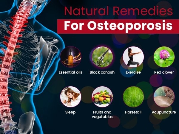
Exercise and dietary optimization form the backbone of nonpharmacologic management you can control immediately. Doing weight-bearing and resistance exercises for 30–45 minutes, three times a week, while gradually increasing the difficulty, can lead to small but important increases in bone mineral density (BMD) of about 1–2% each year and can lower the risk of osteoporosis. Diet should provide adequate protein (about 1.0–1.2 g/kg/day for older adults), calcium from food (dairy, fortified plant milks, and canned sardines), and vitamin D to maintain serum 25(OH)D in the target range many clinicians use (about 30–50 ng/mL). Limiting high-sodium processed foods, excessive caffeine (>3 cups coffee/day), and very-low-calcium vegan diets will reduce bone loss drivers you can modify right away.
Several nutraceuticals have supportive but often modest evidence and should be used with informed expectations. Vitamin K2 (menaquinone MK-7) taken in amounts of about 90–200 mcg per day has been linked in some studies to better bone quality and a lower risk of fractures in postmenopausal women; omega-3 fatty acids (1–2 g per day) and soy isoflavones (50). Collagen peptides (5–10 g/day) can improve some bone-turnover markers and might support matrix quality, though high-quality fracture-endpoint data are limited. Be mindful of interactions—vitamin K interferes with warfarin, and high-dose calcium can affect certain antibiotics and thiazide diuretics—and check baseline labs (serum calcium, creatinine, and vitamin D) before you add supplements.
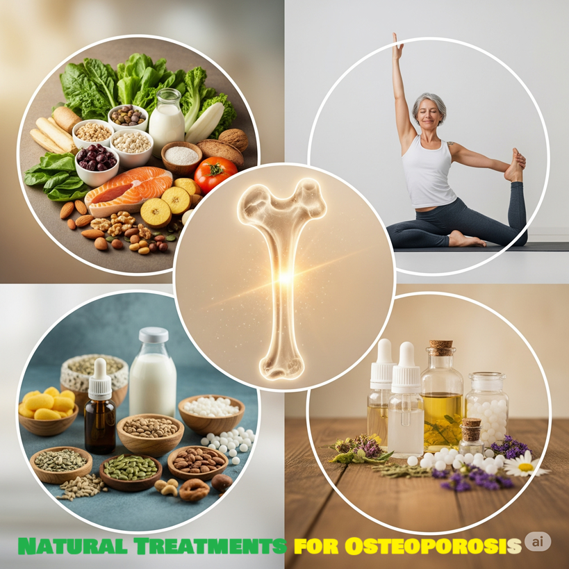
Homeopathic practitioners commonly select remedies based on individualized constitutional assessment rather than on fracture biology: Calcarea carbonica and Calcarea phosphorica are frequently recommended for people with sluggish growth patterns or delayed bone healing; Symphytum (comfrey) is suggested for fracture consolidation; Arnica is used post-trauma to reduce soreness. High-quality randomized control trial prove that homeopathy works for osteoporosis, and reviews show that homeopathic treatments significantly improve bone mineral density or reduce fracture rates compared to a placebo.
1. Calcarea Phosphorica—An Exceptional Remedy for Fragile, Brittle Bones
It is a premier homeopathic remedy for the treatment of osteoporosis. It is mostly administered to people with osteoporosis who possess fragile, soft, and brittle bones that are deformed. The treatment enhances bone density. It is clearly shown when an individual’s hip bone discomfort clearly demonstrates this. Calcarea Phosphorica is advantageous for individuals suffering from back pain attributed to osteopenia. The discomfort may intensify with minimal movement. In certain circumstances, a leftward spinal curvature may be necessary. Occasionally, the lower back may be flexed anteriorly. And finally, it makes broken bones easier to mend.
2. Symphytum Officinale – For the Management of Fractures in Osteoporosis
Symphytum Officinale is the most effective natural treatment for fractures associated with osteoporosis. Commonly referred to as ‘knit bone,’ it facilitates the union of fragmented bone. This therapy effectively facilitates the union of shattered bones by enhancing fibroblast cell activity. This natural remedy significantly alleviates the pricking pain associated with fractures at the injury site. It may also assist in alleviating irritation at the fracture site. This medication may be administered once the fractured bone is immobilized in a cast.
3. Silicea—When Osseous Structures Exhibit Extreme Sensitivity to Palpation
This medication is recommended for instances where the bones exhibit sensitivity and tenderness upon palpation. It is particularly relevant for spinal curvatures predominantly oriented to the right side. It is associated with pain upon contact and movement. In some instances, you may experience a burning feeling in your back. Conditions deteriorate in the open air. Silicea may be used when an individual experiences lower back pain while stooping and rising from a seated position. Silicea is particularly beneficial in cases of pus or fistula formation at the fracture site. Finally, silicea significantly aids in the effective healing of fractured bones and assists in the removal of bone fragments.
4. Calcarea Carbonica—Indicated for weakened spinal and long bones that exhibit curvature.
Calcarea Carbonica is a natural remedy with extensive effects on bone health. It is proven to enhance bone health and fortify fragile and brittle bones. This medication is applicable when the spinal and long bones start to curve due to diminished bone mass. Calcarea Carbonica is effective in alleviating bone discomfort. The discomfort may manifest as dull, pulsing, or stabbing sensations.
5. Mercurius Solubilis – To Alleviate Nocturnal Ostealgia
Mercurius Solubilis is an appropriate medication for alleviating nocturnal bone pain. There is discomfort in the bones, accompanied by a sensation akin to fracture. The discomfort exacerbates upon contact.
6. Phosphorus – For Addressing Spinal Curvature in Osteoporosis
Phosphorus may yield optimal outcomes for individuals experiencing spinal curvature accompanied by a burning sensation in the spine. This natural medicine is effective for alleviating pain and weakness in the spine. The patient experiences severe pain in the spine, like a fracture.
7. Hypericum—For Back Pain Due to Nerve Compression from Injured Vertebrae
Hypericum is an effective natural remedy that can be reliably employed for severe back pain resulting from nerve compression between injured vertebral bones. Hypericum can effectively manage the intense pain and sensitivity in the back following vertebral fractures, which is exacerbated with minimal movement of the neck or arms.
6. Ruta—To Aid in the Repair of Tendons and Ligaments
Accompanied by a Fractured Bone
Ruta is an exceptional natural medication utilized in the management of fractures associated with osteoporosis. When a bone fractures, the adjacent tendons and ligaments also sustain damage. In this domain, Ruta demonstrates its remarkable efficacy in medicine. This natural remedy aids in the repair of ruptured tendons and ligaments. It also aids in the restoration of the compromised bone. This therapy alleviates discomfort, soreness, and stiffness in the vicinity of the broken area, primarily in wrist fractures.
Additional practical guidance: monitor your progress objectively with periodic DEXA scans and, where available, biochemical markers of bone turnover to see whether lifestyle, supplement, or homeopathic additions produce measurable effects; avoid substituting unproven alternatives. Avoid substituting unproven alternatives for prescription drugs if you are at high risk of fractures. Additionally, discuss any over-the-counter or complementary products with your clinician to prevent interactions and gaps in protection that could further increase your fracture risk.
Personalization is key: two patients with the same DEXA T-scores (like a T-score of −2.6 showing osteoporosis) will usually not get the same homeopathic treatment because the practitioner considers your entire background—like your menstrual and reproductive history, past fractures, diet, current medications such as bisphosphonates or denosumab, and your emotional state—care with evidence-based prevention measures: aim for total calcium intake near 1,000–1,200 mg/day and a 25(OH)D target above 30 ng/mL, continue weight-bearing exercise, and maintain fall-prevention strategies. Check your bone density with DEXA every 1–2 years as your doctor advises so that any additional homeopathic treatment can be assessed based on changes in bone strength and fracture risk, not just on how you feel.
Professional Guidance: Finding a Homeopath
Find a practitioner who has formal training and experience in treating long-term bone issues; check for certification from well-known organizations like the Faculty of Homeopathy (UK) or the Council for Homeopathic Certification (US), or ensure they have at least three years of advanced clinical training and supervised practice.
Ask prospective homeopaths how many patients with osteoporosis or osteopenia they have treated, and request case summaries or anonymized outcomes. A clinician who regularly records initial DEXA scores, serum 25(OH)D levels, and follow-up functional measures is collaborating with your primary care doctor, endocrinologist, or orthopedic surgeon to ensure that regular check-ups (like lab tests, imaging, and medication management) can proceed smoothly—this is especially important if you are taking bone-specific medications like bisphosphonates, SERMs, or partial homeopathic consultation to be comprehensive: A typical consultation lasts 60–90 minutes, during which the practitioner will take a detailed medical history, including information on fractures and medications; review your most recent DEXA report and lab results (calcium, vitamin D, and PTH); and assess lifestyle factors such as dietary sodium, protein intake, alcohol consumption, smoking habits, and exercise routines. Follow-up visits tend to be shorter—15–30 minutes—at intervals of 4–12 weeks initially to allow the remedy response to declare itself; many homeopaths adjust potency or change remedies based on symptom shifts rather than on a predetermined schedule. Typical fees vary by region but expect an initial visit between $275 and $300 and follow-ups of $90–$150; some clinics offer integrated care packages that include coordination with physiotherapy or nutritional counseling focused on an osteoporosis diet that avoids excess sodium and phosphates and emphasizes calcium-rich whole foods.
Watch for red flags during your search: any practitioner who advises you to stop prescribed osteoporosis medications without consultation with your treating physician or who promises a cure for established osteoporosis (T-score ≤ −2.5 or previous fragility fracture) should be avoided. Make sure the homeopath knows important signs of bone health—like how to read a DEXA report, understand a FRAX score, and the importance of serum 25(OH)D and PTH—and that they will send you back to regular doctors if you have serious symptoms like sudden severe back pain that could mean a vertebral fracture. Health options are increasingly available, but you should ensure that remote practitioners still require recent DEXA and lab results and commit to ongoing communication with your medical team.
Don’t forget that a homeopathic practitioner is first of all a medical doctor. If a homeopath is not a medical doctor, he is not a homeopath.
Sample questions to bring to an initial meeting: “How many osteoporosis patients have you managed, and can you share anonymized outcomes?” “How will you coordinate with my endocrinologist or primary care physician?” “Which remedies do you commonly use for bone fragility and what potencies/frequencies do you typically select?” “How do you measure treatment response—symptoms, functional tests, DEXA interval?” These direct queries will help you assess whether the homeopath’s methodology meets your requirements for integrated, measurable, and safe care as you pursue strategies to strengthen your bones.
Building Resilience: Proven Strategies to Prevent Osteoporosis
Lifestyle Modifications: Exercise, Diet, and Habit Changes
Targeted exercise programs provide the most significant, clinically proven benefits for improving bone density and preventing falls. Combine weight-bearing aerobic activities like brisk walking, stair climbing, and jogging with strength training using free weights, resistance machines, or bodyweight exercises two to three times a week; studies show that doing both types of exercise together can lead to noticeable increases in bone density in the lower back and hips of about 1–3% each year in adults. Add short bouts of higher-impact moves (jumping, heel drops, or hopping for those without contraindications) 2–3 times weekly to stimulate bone formation at the hip and tibia, and use balance and gait training—Tai Chi, single-leg stands, and dynamic balance drills—to reduce fall risk by up to roughly 30% in older adults, according to fall-prevention trials.
Nutrition directly supplies the building blocks for bone remodeling: aim for a total calcium intake of about 1,000–1,200 mg daily depending on your age and sex, and target serum 25(OH)D in a range clinicians commonly use (roughly 20–30 ng/mL, with many treating toward the upper end for fracture prevention). Prioritize protein intake at approximately 1.0–1.2 g/kg/day in older adults to support bone matrix and muscle mass—clinical studies link higher protein with lower fracture risk when calcium intake is adequate. Cut back on dietary factors that accelerate calcium loss or impair bone quality: keep sodium moderate (excess adds to urinary calcium loss), limit daily alcohol to well below two standard drinks (higher consumption increases fracture risk), and avoid very high caffeine intake without compensating calcium; also reduce excessive intake of foods high in oxalates if you rely on plant sources for calcium because absorption can be affected.
Daily habits and home modifications sustain gains from exercise and diet by lowering injury risk and maintaining functional independence. You can lower fall probability by installing grab bars, improving lighting, removing loose rugs, and using non-slip footwear—intervention studies in community-dwelling older adults show environmental changes plus exercise halve fall-related hip fractures in some cohorts. Quitting smoking yields direct benefits for bone health: smokers experience higher rates of osteoporosis and delayed fracture healing, so stopping smoking leads to measurable reductions in future fracture risk over time.. Finally, coordinate with your clinician regarding supplements and medications: if you have a limited dietary intake or inadequate sun exposure, taking a vitamin D supplement (typically 800–1,000 IU/day for older adults, tailored by lab results) and calcium supplementation as necessary can be safe adjuncts while you pursue lifestyle changes.
Regular Screenings: Staying Ahead of Bone Health
Bone-mineral density assessment by dual-energy X-ray absorptiometry (DXA) remains the cornerstone of screening; you should plan for a baseline DXA at the guideline-recommended ages—women at 65 and men commonly at 70, or earlier if you have risk factors such as glucocorticoid use, prior fragility fracture, low body weight, or hypogonad. Understanding T-scores helps determine your bone health: a T-score between −1.0 and −2.5 means you have osteopenia, and a score of −2.5 or lower means you have osteoporosis. This increases monitoring frequency. Use the FRAX 10-year fracture risk calculator. Any US-based guidelines recommend considering treatment when the 10-year hip fracture risk reaches ≥3% or when the major osteoporotic fracture risk is ≥20%. osteoporotic fracture risk is ≥20%.
Beyond DIn addition to DXA scans, do specific tests to find other causes and hidden fractures—check levels of serum 25(OH)D, calcium, creatinine, and thyroid-stimulating hormone, and for men, also lookal fracture assessment (VFA) using DXA or conventional lateral spine radiographs can detect asymptomatic compression fractures in up to 20% of older adults who are screened; discovering even one vertebral fracture significantly increases the estimated fracture risk and often shifts management from solely prevention to active therapy. If you are on osteoporosis medications, plan follow-up DXA at intervals tailored to therapy and risk: Many clinicians initially repeat imaging every 1–3 years to document the response and then every 2–3 years once the condition is stable, while also selectively tracking markers such as bone turnover tests to gauge adherence and biological response.
You should integrate fall-risk screening into every visit—simple tools like the Timed Up and Go (TUG) test, gait speed, and home-safety checklists identify more. Think about referring the patient to an endocrinologist or a metabolic bone clinic if they are experiencing ongoing bone loss despite treatment, have unusual fractures, or if lab tests show multiple secondary causes; getting a team of specialists involved early helps improve diagnosis and customize treatment length, especially when considering breaks from medication or switching between different types of bone-strengthening drugs. Antiresorptives and anabolic agents play a significant role.
Conclusion
Summing up, osteoporosis is a progressive reduction in bone density and microarchitectural integrity that increases your risk of fractures; it arises from a combination of biological factors (age-related bone loss, hormonal changes such as menopause, and genetic predisposition), nutritional deficits (longstanding low calcium and vitamin D intake), lifestyle contributors (sedentary behavior, smoking, and excessive alcohol), and certain medications (long-term glucocorticoids and some anticonvulsants). You will often notice subtle signs such as losing height, having a hunched back, unexplained back pain, or breaking bones easily from minor falls, but a clear diagnosis requires specific tests—usually DEXA bone mineral density testing and certain blood tests—and distinguishing it from similar conditions (osteopenia is when bone mass is lower than normal but not low enough to be osteoporosis; osteomalacia is when bones don’t mineralize properly due to a lack of vitamin D). both you and your clinician to interpret tests and prioritize interventions that are tailored to your specific risk profile. Tailor prevention and dietary management are essential for maintaining management and are central to preserving your skeletal health over the long term. Focus on eating foods that are high in calcium, enough protein, and vitamin D from both food and safe sun exposure or supplements when needed; include leafy greens, dairy or fortified alternatives, oily fish, and legumes, and make sure to have protein in your meals regularly to help support bone health.actors that accelerate calcium loss or impair bone formation—high-salt diets, excessive caffeine, frequent cola intake, very-high-phosphate processed foods, and heavy alcohol use—and avoid crash diets that compromise nutrient intake. Combine nutrition with regular weight-bearing and resistance exercises, balance and mobility training to reduce fall risk, smoking cessation, and home modifications to prevent falls. You should also engage in age- and risk-appropriate screening and follow-up bone density testing so that emerging osteopenia or osteoporosis can be managed early.
Treatment options span proven pharmacological treatment options and include effective medications, lifestyle changes, and alternative methods that some people use for symptom relief. Bisphosphonates, SERMs (like raloxifene), RANKL inhibitors (denosumab), and anabolic agents (teriparatide, abaloparatide, and romosozumab)—are used to reduce fracture risk and are selected based on your fracture history, bone density, comorbidities, and tolerance; calcium and vitamin D supplementation often accompanies these therapies and monitoring for side effects and long-term Natural measures that can strengthen your overall bone health include targeted nutrition, structured exercise programs, fall-prevention strategies, and physiotherapy aimed at optimizing posture and function. Homeopathy is used by some individuals, and homeopathic practitioners may suggest remedies such as Calcarea carbonica, Symphytum or Arnica tailored to your symptoms’ pattern, but high-quality evidence for clinically meaningful effects on bone density is limited; any complementary approach should be coordinated with your medical team so you retain effective, evidence-based fracture prevention while addressing your broader health needs.
FAQ

Q: What is osteoporosis?
A: Osteoporosis is a systemic skeletal disorder characterized by low bone mass and deterioration of bone microarchitecture, which increases bone fragility. Osteoporosis is often asymptomatic until a fragility fracture occurs, which commonly affects the vertebrae, hip, or wrist (commonly vertebral, hip, or wrist). Bone density is typically measured by dual-energy X-ray absorptiometry (DEXA) and reported as a T-score to guide diagnosis and management.
Q: What causes osteoporosis?
Bone loss occurs due to an imbalance between the processes of bone resorption and bone formation.
Two primary causes of osteoporosis include age-related bone loss and hormonal changes, particularly estrogen deficiency that occurs after menopause. Secondary causes of bone loss include long-term glucocorticoid use, hyperthyroidism, hypogonadism, malabsorption (such as celiac disease), chronic kidney disease, certain anticonvulsants, aThe major risk factors for osteoporosis include older age, female sex, a family history of fractures, low body weight or small frame, low intake of calcium and vitamin D, a sedentary lifestyle, smoking, excessive alcohol consumption, and prolonged use of steroids or other medications that impact bone health.
Q: What are common symptoms and signs of osteoporosis, and how does it differ from osteopenia and osteomalacia?
A: Osteoporosis is often asymptomatic until a fracture occurs. Symptoms/signs may include acute or chronic back pain from vertebral compression fractures, loss of height, kyphosis (stooped posture), and Osteopenia is defined as lower-than-normal bone density that does not meet the T-score threshold for osteoporosis, which ranges from −1.0 to −2.5; this condition indicates an increased risk of fractures but involves milder bone loss. Osteomalacia carries an increased risk but results in milder bone loss. Osteomalacia is a defect in bone mineralization usually caused by severe vitamin D deficiency or phosphate disorders, presenting with bone pain, muscle weakness, and looser (pseudofracture) lines; laboratory findings (low 25‑OH vitamin D, low serum phosphate, and elevated alkaline phosphatase) help distinguish it from osteoporosis.
Q: How is osteoporosis diagnosed?
A: Diagnosis is based on bone mineral density measurement by DEXA. A T-score ≤ −2.5 at the lumbar spine, femoral neck, or total hip defines osteoporosis. A T-score between Clinical tools, such as FRAX, estimates the 10-year fracture risk and assists in determining treatment options. Imaging (X-ray) may show fractures or vertebral compression. Laboratory tests (calcium, phosphate, 25‑OH vitamin D, PTH, TSH, renal function, CBC, and markers of bone turnover) are used to identify secondary causes and guide management.
Q: What should someone with osteoporosis avoid eating, and what dietary pattern supports bone health?
A: Limit or avoid excess alcohol, heavy caffeine intake, and frequent cola-type sodas (high phosphoric acid) because these can impair bone health. Avoid very high-salt diets and excessive preformed vitamin A (retinol) supplements, which may increase fracture risk. A bone-supportive diet emphasizes adequate calcium (dairy, fortified plant milks, leafy greens, canned oily fish with bones), sufficient protein, vitamin D (fatty fish, fortified foods, sensible sun exposure, supplements if needed), magnesium, and vitamin K. Spread calcium intake evenly across meals to optimize absorption, and consult a clinician about supplements if dietary intake is insufficient.
Q: What medical treatments and nonpharmacologic measures are used for osteoporosis?
A: The main medication options for people at high risk of fractures include bisphosphonates (like alendronate, risedronate, and zolesuch as alendronate, risedronate, and zoledronic acid), denosumab, and for some patients, selective estrogen receptor modulators like raloxifene or hormone replacement therapyequate calcium and vitamin D, individualized fall prevention strategies, weight-bearing and resistance exercises, smoking cessation, and limitations on alcohol consumption. Medication choice depends on fracture risk, comorbidities, preferences, and contraindications; potential adverse effects (esophageal irritation, osteonecrosis of the jaw, atypical femur fracture) and monitoring should be discussed with a provider.
Q: What is the role of homeopathy and natural remedies in managing osteoporosis?
A: Many patients use homeopathic remedies, and high-quality clinical evidence demonstrates their ability to prevent fractures. Commonly cited homeopathic remedies include Calcarea carbonica, Calcarea phosphorica, and Symphytum, among others; however, these remedies should not replace evidence-based medical treatment for individuals who are at significant risk of fractures. Natural measures that have supportive evidence include regular weight-bearing and resistance exercise, adequate dietary calcium and vitamin D intake, maintaining a healthy body weight, and addressing secondary causes such as treating vitamin D deficiency (e.g., treating vitamin D deficiency). Discuss any complementary therapies or supplements with a healthcare professional to avoid interactions and ensure comprehensive fracture-risk management.
Need Treatment for Osteoporosis?
Sadly, there’s no cure for osteoporosis. This condition is very painful and can prevent you from doing the most routine tasks. However, with proper care and treatment, you can slow down the progress of osteoporosis and even promote bone growth. With conventional treatments and drugs, there’s always the danger of side effects. Additionally, they don’t work on everyone and might be too aggressive for some. If you want a more natural approach, you can try the above-mentioned natural remedies for osteoporosis. They offer holistic treatment without any side effects. If you still don’t see results, you must talk to your doctor immediately. The Philadelphia Holistic Clinic is the center for bone and joint diseases. To schedule an initial evaluation at the center, please contact us or use our online application. Choose “Book an Appointment” on the top menu and follow simple instructions.
You must be logged in to post a comment.